Showing posts with label biolog science. Show all posts

Sunday, May 1, 2011

Gene Therapy


The best way of treating a cancer would be to find a way of genetically modifying the tumour cells, correcting the genetic defect. This technique is known as ‘gene therapy’, It is regarded as a potential revolution in medicine because gene therapy is aimed at treating or eliminating causes of disease, whereas most current drugs treat the symptoms.


Gene therapy for cancer accounts for the majority of gene therapy clinical trials. Targets for
this include replacement of tumor suppressor genes, ‘‘suicide genes’’ to activate prodrugs, antiangiogenic gene therapy, cytokine-based gene transfer, and delivery of drug resistance genes to hematopoietic stem cells to protect them from the bone marrow toxicity of chemotherapeutic agents.

Suicide gene therapy approaches include transfection of tumor cells with herpes simplex
virus-thymidine kinase (HSV-TK) to activate the prodrug ganciclovir and cytosine deaminase
to convert the nontoxic compound 5-fluorocytosine into cytotoxic 5-fluorouracil .

Suicide gene clinical trials have been carried out in prostate cancer, mesothelioma, and glioblastoma. Cytokine gene therapy approaches have included gene transfer via tumor-homing lymphocytes bearing genes encoding interleukins (IL-1B, IL-2, IL-4, IL- 12), GM-CSF, and interferon-g (IFN-g). Clinical trials include IL-12 delivered by a vaccinia virus vector for mesothelioma and GM-CSF and IFN-g delivered in retroviral vectors for melanoma. Some clinical responses have been observed in these trials with small numbers of patients.

The tumor suppressor gene p53 is the most commonly mutated gene in human cancers.
Hence, it is a good target for gene replacement  therapy. Re-expression of p53 in human colon cancer cell lines bearing a mutated gene inhibits tumor cell proliferation. In a murine model of p53-mutated colon cancer, injection of an adenoviral vector encoding the WT p53 gene into tumors resulted in tumor regression and enhanced survival . Clinical trials with p53 gene replacement have been initiated for a number of cancers including colon and
head and neck cancers. In general, the procedures were well tolerated. The main side effects were fever and transient liver enzyme abnormalities. One key question for this and other gene therapy approaches to cancer is how many cells in a tumor need to be transfected to get a therapeutic effect? There is some evidence for ‘‘bystander’’
effects from p53 transfection, probably due to an antiangogenesis effect.620 Other potential tumor suppressor gene targets for which there is at least preclinical demonstration of efficacy are PTEN, E-cadherin, C-CAM, BRCA-1, and pHyde.

TUMOUR SUPPRESSOR GENES


Tumour suppressor genes prevent excessive growth of a cell; the most well known ones are p53 and the retinoblastoma (Rb) gene.

Retinoblastoma Gene

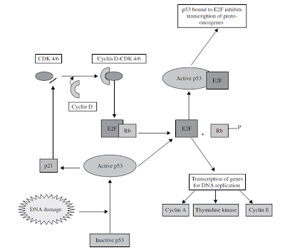
Retinoblastoma gene is involved in the G1 checkpoint in the following way. It binds to a family of transcription factors known as the E2F family, thereby repressing their transcription of E2F-responsive genes, such as thymidine kinase (TK), needed for DNA replication, and cyclin E and A, needed for cell cycle progression. Rb is activated when cyclin D forms a complex with CDK4/6 (cyclin D/CDK4/6, hence making it active) this in turn phosphorylates Rb, which allows E2F to be released

p53

The p53 protein is essential for protecting us against cancer. More than half of human cancers have p53 mutations and therefore no functioning p53. p53 works by sensing DNA damage and halting the cell cycle (Figure 4.2). This is essential, because if DNA is damaged but still replicated in S phase, it could eventually manifest in the form of a protein mutation. By halting the cell cycle at the G1 checkpoint, this can be prevented. So how does this process work? Again, it comes back to the involvement of CDKs. First, in response to a variety of stress signals,

for example DNA damage, p53 switches from an inactive state to an active state. It then triggers transcription of the gene for p21, which is a CDK inhibitor. Because active CDKs are needed to progress through the cell cycle, an inactive CDK will cause the cycle to halt. The p53 protein is also involved at the G2 checkpoint in cases, for example, where DNA has been synthesized incorrectly. At this checkpoint, p53 binds to E2F (see Section ‘Retinoblastoma gene’) and prevents it from triggering transcription of proto-oncogenes, for example c-myc and c-fos, which are required for mitosis Proto-oncogenes are important promoters of normal cell growth and division; however, if they become mutated they are known as oncogenes and can have a detrimental effect. A single oncogene cannot cause cancer by itself but it can cause the cell cycle to lose its inhibitory controls, thereby increasing the rate of mitosis. When a cell loses control over mitosis, it can be the beginning of the pathway leading to the development of cancer

Tuesday, October 26, 2010

How To Make a DNA Model Using Candy...


Making DNA models can be informative, fun, and in this case tasty. Here you will learn how to construct a DNA model using candy. But first, what is DNA? DNA is a nucleic acid that contains the genetic information for the reproduction of life. Its shape is that of a double helix and its appearance is somewhat of a twisted ladder or spiral staircase. DNA is composed of nitrogenous bases (adenine, cytosine, guanine and thymine), a five-carbon sugar (deoxyribose), and a phosphate molecule.

Nucleic Acids


                                                                             Nucleic acids allow organisms to transfer genetic information from one generation to the next. There are two types of nucleic acids: deoxyribonucleic acid, better known as DNA and ribonucleic acid, better known as RNA.

When a cell divides, its DNA is copied and passed from one cell generation to the next generation. DNA contains the "programmatic instructions" for cellular activities. When organisms produce offspring, these instructions, in the form of DNA, are passed down. RNA is involved in the synthesis of proteins. "Information" is typically passed from DNA to RNA to the resulting proteins.
Nucleic acids: Nucleotides
Nucleic acids are composed of nucleotide monomers. Nucleotides have three parts:
  • A Nitrogenous Base
  • A Five-Carbon Sugar
  • A Phosphate Group
Similar to what happens with protein monomers, nucleotides are linked to each other through dehydration synthesis. Interestingly, some nucleotides perform important cellular functions as "individual" molecules, the most common example being ATP.
Polynucleotides
In polynucleotides, nucleotides are joined to one another by covalent bonds between the phosphate of one and the sugar of another. These linkages are called phosphodiester linkages

The Cell-Cell Structure...


Life is both wonderful and majestic. Yet for all of its majesty, all organisms are composed of the fundamental unit of life, the cell. The cell is the simplest unit of matter that is alive. From the unicellular bacteria to multicellular animals, the cell is one of the basic organizational principles of biology. Let's look at some of the components of this basic organizer of living organisms.
Eukaryotic Cells and Prokaryotic Cells
                                                                                                          There are two primary types of cells: eukaryotic cells and prokaryotic cells. Eukaryotic cells are called so because they have a true nucleus. The nucleus, which houses DNA, is contained within a membrane and separated from other cellular structures. Prokaryotic cells however have no true nucleus. DNA in a prokaryotic cell is not separated from the rest of the cell but coiled up in a region called the nucleoid.

                                                                                                        As organized in the Three Domain System, prokaryotes include archaeans and bacteria. Eukaryotes include animals, plants, fungi and protists. Typically, eukaryoitc cells are more complex and much larger than prokaryotic cells. On average, prokaryotic cells are about 10 times smaller in diameter than eukaryotic cells.

Eukaryotes grow and reproduce through a process called mitosis. In organisms that also reproduce sexually, the reproductive cells are produced by a type of cell division called meiosis. Most prokaryotes reproduce through a process called binary fission. During binary fission, the single DNA molecule replicates and the original cell is divided into two identical daughter cells.

Both eukaryotic and prokaryotic organisms get the energy they need to grow and maintain normal cellular function through cellular respiration. Cellular respiration has three main stages: glycolysis, the citric acid cycle, and electron transport. In eukaryotes, most cellular respiration reactions take place within the mitochondria. In prokaryotes, they occur in the cytoplasm and/or within the cell membrane.
The Cell-Cell Structure
There are also many distinctions between eukaryotic and prokaryotic cell structure. The following table compares the cell structures found in a typical prokaryotic cell to those found in a typical animal eukaryotic cell.
Cell Structure Comparison
Eukaryotic and Prokaryotic Cell Structure

Cell Structure
Prokaryotic Cell
Typical Animal Eukaryotic Cell
Cell Wall
Yes
No
Centrioles
No
Yes
Chromosomes
One long DNA strand
Many
Cilia or Flagella
Yes, simple
Yes, complex
Endoplasmic Reticulum
No
Yes (some exceptions)
Golgi Complex
No
Yes
Lysosomes
No
Common
Mitochondria
No
Yes
Nucleus
No
Yes
Peroxisomes
No
Common
Cell Membrane
Yes
Yes
Ribosomes
Yes
Yes


Animal Tissues: Epithelial Tissue


What are Tissues?
                                 The word tissue is derived from a Latin word meaning to "weave." Cells that make up tissues are sometimes "woven" together with extracellular fibers.

Likewise, a tissue can sometimes be held together by a sticky substance that coats its cells.

There are four main categories of tissues: epithelial, connective, muscle and nervous. Let's take a look at epithelial tissue.
Epithelial Tissue
Epithelial tissue covers the outside of the body and lines organs and cavities. The cells in this type of tissue are very closely packed together and joined with little space between them.

     With a tightly packed structure we would expect epithelial tissue to perhaps serve some type of barrier and protective function and that is certainly the case.

Epithelial tissue helps to protect organisms from microorganisms, injury, and fluid loss.

In an epithelium, the free surface is usually exposed to fluid or the air while the bottom surface is attached to a basement membrane.
Classifying
Epithelia are commonly classified based on the shape of the cells on the free surface, as well as the number of cell layers. Sample types include:

Simple Epithelium: A simple epithelium has a single layer of cells.

Stratified Epithelium: A stratified epithelium has multiple layers of cells.

Likewise, the shape of the cells on the free surface can be:

Cuboidal

Analogous to the shape of dice.

Columnar

Analogous to the shape of bricks on an end.

Squamous

Analogous to the shape of flat tiles on a floor.


By combining the terms for shape and layers, we can derive epithelial types such as stratified squamous epithelium or simple columnar epithelium.
Animal Tissue Types
To learn more about animal tissues, visit:
  • Connective Tissue
  • Muscle Tissue
  • Nervous Tissue
  • Nervous Tissue - Glial Cells
Share Your Opinions
What do you think? Have questions about animal tissue types or epithelial tissue? Come on over to the Biology Forum and share your thoughts, opinions, and feelings...........